Narragansett Bay Adult Oyster DNA Extractions - Part 3
DNA Extraction of Adult Oyster Tissue - Part 3
~10 wild adult oysters were collected from 5 populations in Narragansett Bay
- Narrow River
- Green Hill Pond
- Barrington River
- Kickemuit
- Mary C. Donovan Marsh
Mantle and gill tissue were dissected from the oysters. Dissection protocol can be accessed here.
DNA Extractions
Completed on January 25, 2021
Zymo Research Quick-DNA Miniprep Plus used for DNA extractions of adult oyster tissue
- Pull samples out of -80 freezer and put on ice
- NAR_5
- NAR_8 - mantle and gill
- KIC_3
- MCD_2
- MCD_5
- Pull Proteinase k out of upright -20 freezer in Puritz Zymo Reagents box and put on ice
- In original sample tube, add 90 ul of Type II DI water and 10 ul of 10X PBS (Phosphate Buffered Saline) solution to create 1X PBS solution soak
- Vortex and spin down in mini centrifuge
- Let soak for 5-10 minutes while labeling new 1.5-ml tubes
- Ended up being closer to 15-20 minutes
- Pull out Blue Solid Tissue Buffer from Zymo Research Quick DNA Miniprep kit
- In newly labeled 1.5-ml tubes, add 95 ul of nuclease-free water, 95 ul of solid tissue buffer, and 10 ul of Proteinase k
- Vortex and spin down in mini centrifuge
- Sterilize forceps with 10% bleach, Type II DI water, and 70% EtOH
- Transfer tissue pieces from PBS tube to respective 1.5-ml tube
- Sterilize forceps before each sample
- Vortex samples for 10 seconds and spin down in mini centrifuge
- Put in thermomixer at 55 deg C at 600 rpm for 45 minutes
- Using half speed from what was done with previous samples
- Tissue was not fully solubilized after 45 minutes, so increased speed to 1000 rpm for 30 more minutes
- Once tissue is fully solubilized, place all samples in tabletop centrifuge and spin at 13000 rcf for 2 minutes
- 1 minute longer than previous samples
- Make new set of labeled 1.5-ml tubes
- Pipette all supernatant (200 ul) to labeled tubes
- Make 1.5 ml tube of 10 mM Tris HCl pH 8 and place in thermomixer at 70 deg C
- Set up tubes for extraction - 1 yellow spin column inside a collection tube for each sample - label lid of spin column
- Get liquid waste beaker from sink near -80 freezers
- Add 2 parts volume (400 ul) of Genomic Binding buffer to each tube
- Vortex for 5 seconds and spin down in mini centrifuge
- Add 400 ul of sample to their labeled yellow spin column
- Centrifuge spin columns at 13000 rcf for 1 minute
- Pour off flow through in liquid waste beaker
- Put spin columns in same collection tubes
- Add remaining liquid (200 ul) from each sample to labeled yellow spin column
- Centrifuge spin columns at 13000 rcf for 1 minute
- Pour off flow through in liquid waste beaker
- Transfer spin columns to new collection tubes and discard of old collection tubes
- Add 400 ul of DNA pre-wash buffer to each spin column
- Centrifuge at 13000 rcf from 1 minute
- Pour off flow through in liquid waste beaker
- Place spin columns in same collection tubes
- Add 700 ul of g-DNA wash buffer to each spin column
- Centrifuge spin columns at 13000 rcf for 1 minute
- Pour off flow through in liquid waste beaker
- Put spin columns in same collection tubes
- Add 200 ul of g-DNA wash buffer to each spin column
- Centrifuge spin columns at 13000 rcf for 1 minute
- Make final 1.5 ml tubes - label lid with sample id, elution #, and DNA; label side with initials, date of extraction, sample id, elution #, DNA, and C. virginica
- Make 2 tubes for each sample - splitting the two elutions into two separate tubes
- Transfer spin columns to first set of labeled 1.5 ml tubes
- Pour off flow through in liquid waste beaker and discard collection tubes
- Take warmed 10 mM Tris HCl pH 8 out of thermomixer
- Add 50 ul of warmed 10 mM Tris HCl pH 8 to each spin column by dripping directly over the filter without touching it
- Incubate for 5 minutes
- Place tubes in centrifuge with all the lids of the 1.5 ml tubes facing the same direction and centrifuge at MAX speed for 1 minute
- Take tubes out - DO NOT pour off liquid, keep in tube
- Transfer spin columns to second set of labeled 1.5-ml tubes, add 50 ul of warmed 10 mM Tris HCl pH 8 to each spin column by dripping directly over filter without touching it
- Put first set of 1.5-ml tubes with the first elution on ice
- Incubate for 5 minutes
- Centrifuge at max speed for 1 minute
- Take out spin columns at discard
- Make labeled PCR strip tubes - 1 for each elution = 2 per sample
- Add 2 ul of DNA to the respective PCR tube for agarose gel
- 1 ul will be taken directly from 1.5-ml tube for Qubit
47 ul remaining in each 1.5 ml sample tubes
Qubit dsDNA BR assay
The captured pools were quantified following Qubit protocol for BR DNA
| Sample | Avg ng/μl |
|---|---|
| Std 1 | 174 RFU |
| Std 2 | 19572 RFU |
| NAR_5 E1 | 45.5 |
| NAR_5 E2 | 6.51 |
| NAR_8 M E1 | 72.3 |
| NAR_8 M E2 | 9.29 |
| NAR_8 G E1 | 60.6 |
| NAR_8 G E2 | 8.55 |
| KIC_3 E1 | 30.8 |
| KIC_3 E2 | 3.45 |
| MCD_2 E1 | 35.5 |
| MCD_2 E2 | 3.41 |
| MCD_5 E1 | 30.4 |
| MCD_5 E2 | 2.97 |
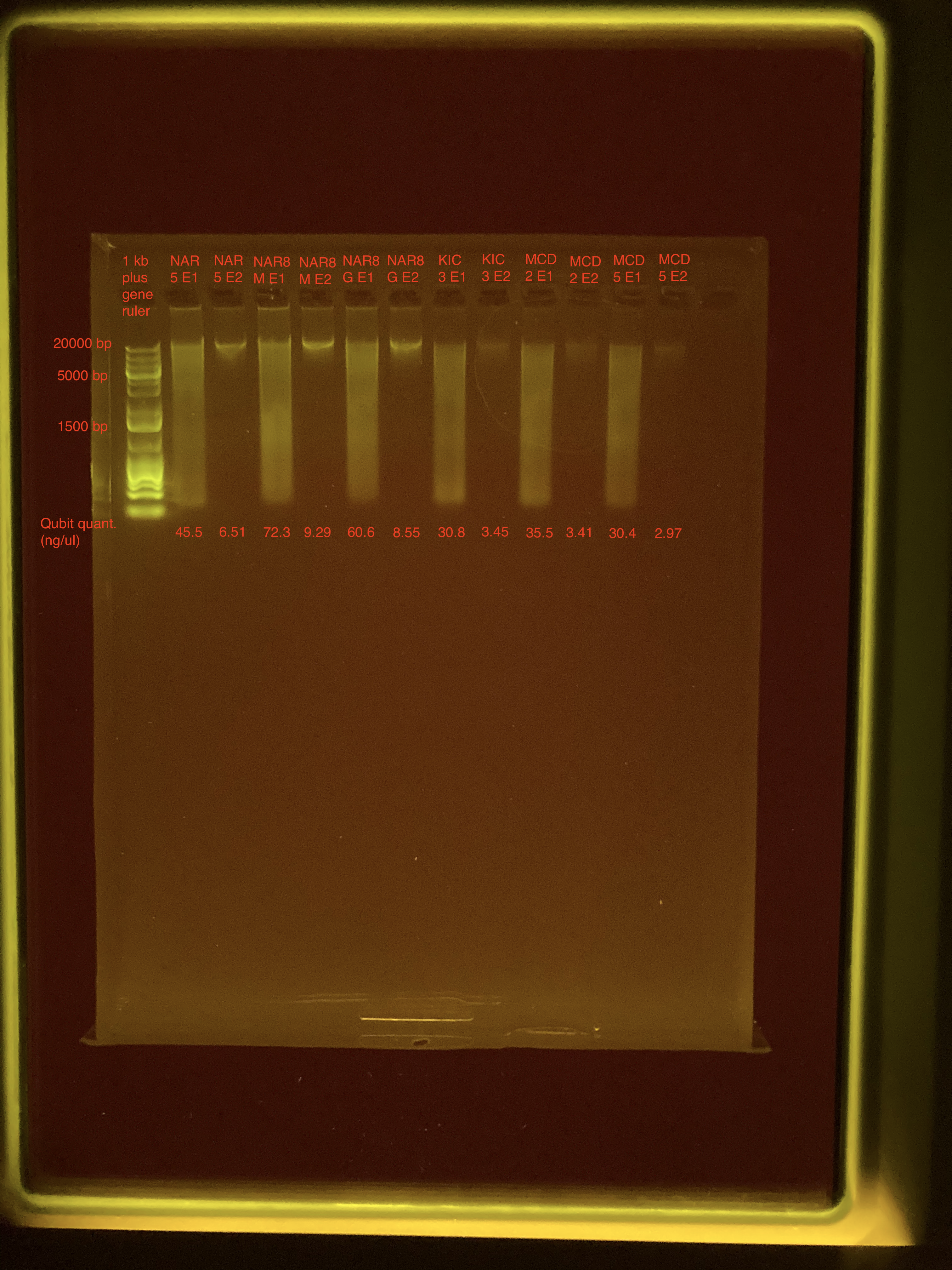
Much smaller quantities of DNA in the second elution, these have the larger fragments.
Agarose Gel Electrophoresis
The DNA quality and size were assessed following Agarose Gel Protocol for a small 1% gel.
- Only 2 ul of DNA for each sample were used
- Did not use diluted gelgreen this time
- Used a different loading dye - Purple loading dye versus Tritrack

The second elution of each sample has the high quality DNA but in low quantity.
Plan for next set of extractions:
- Do PBS soak again
- Maybe dissect more gill tissue
- When separating the two elutions, use a smaller volume for elution 1 and larger volume for elution 2(?)
